Diagnosing mesothelioma requires a systematic and thorough approach that combines multiple medical techniques to accurately identify and confirm the presence of this rare cancer.
Medical professionals employ a range of diagnostic methods to detect and analyze potential mesothelioma cases, ensuring patients receive the most precise evaluation possible.
Initial Physical Examination
The initial physical examination serves as a critical first step in identifying potential mesothelioma indicators.
Doctors conduct a comprehensive assessment that focuses on gathering detailed medical history, particularly regarding asbestos exposure, and performing a thorough physical evaluation to detect early signs of the disease.
Here are the key elements physicians examine during an initial physical assessment:
- Medical History: Detailed review of patient’s occupational background, potential asbestos exposure, and family medical history
- Symptom Evaluation: Comprehensive assessment of mesothelioma symptoms like chest pain, shortness of breath, and unexplained weight loss
- Physical Inspection: Careful examination of chest and abdominal regions for signs of fluid buildup or abnormal masses
- Risk Factor Analysis: Identification of potential risk factors associated with asbestos exposure and cancer development
The physical examination provides physicians with initial insights that guide further diagnostic procedures, helping them determine the most appropriate next steps in identifying potential cancer cells.
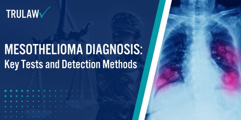
Advanced Imaging Techniques
Advanced imaging techniques play a pivotal role in detecting and analyzing potential mesothelioma locations and characteristics.
These sophisticated diagnostic tools allow medical professionals to obtain detailed internal views that help identify suspicious areas requiring further investigation.
Physicians utilize the following advanced imaging methods:
- Computed Tomography (CT) Scans: High-resolution cross-sectional images that reveal detailed views of chest and abdominal regions
- Positron Emission Tomography (PET) Scans: Radioactive tracer-based imaging that helps identify potential cancer activity
- Magnetic Resonance Imaging (MRI): Detailed soft tissue imaging that provides comprehensive views of potential tumor locations
- X-rays: Initial screening method that can identify fluid buildup or initial signs of pleural mesothelioma
These advanced imaging techniques enable doctors to create a comprehensive map of potential peritoneal mesothelioma locations, guiding subsequent diagnostic procedures like biopsies.
By combining multiple imaging methods, physicians can develop a more accurate understanding of the disease’s progression and potential impact on tissue samples and lymph nodes.
Biopsy Procedures and Their Significance
Biopsy procedures represent the most definitive method for diagnosing mesothelioma, providing physicians with direct tissue evidence of the disease.
These procedures allow doctors to extract and examine tissue samples that reveal critical information about the cancer’s characteristics and progression.
Here are the primary biopsy methods used to diagnose mesothelioma:
- Thoracoscopy: A minimally invasive surgical procedure that allows direct visualization of the chest cavity and collection of tissue samples for malignant pleural mesothelioma diagnosis.
- Surgical Biopsy: An invasive method used when other biopsy techniques cannot obtain sufficient tissue samples for accurate analysis.
- Needle Biopsy: A less invasive technique that uses fine needles to extract small tissue samples from suspicious areas.
- Laparoscopy: An endoscopic procedure particularly effective for diagnosing peritoneal mesothelioma by examining the abdominal cavity.
The selection of a specific biopsy method depends on the tumor’s location, size, and accessibility.
Each technique provides pathologists with essential tissue samples that help determine the precise nature of the mesothelioma cells and guide subsequent treatment strategies.
Pathological Analysis and Laboratory Tests
Pathological examination plays a critical role in confirming a mesothelioma diagnosis by providing detailed insights into the cellular characteristics of the disease.
Specialized mesothelioma pathologists analyze tissue samples with extraordinary precision to identify unique markers and cellular structures.
Key laboratory tests used in mesothelioma diagnosis include:
- Immunohistochemical Markers: Advanced testing that identifies specific protein markers characteristic of mesothelioma tumors.
- Biomarker Testing: Blood tests that detect specific molecular indicators associated with mesothelioma development.
- Cell Type Identification: Microscopic analysis to determine whether the cancer has spread and classify the specific mesothelioma cell type.
- Fluid Cytology: Examination of fluid samples, such as pleural effusion, to detect cancer cells.
Laboratory tests complement biopsy procedures by providing additional context about the disease’s progression and potential treatment responses.
These sophisticated analytical methods enable physicians to develop targeted treatment plans that address the unique characteristics of each patient’s mesothelioma.